Case Overview
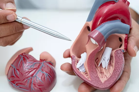
A congenital heart defect (CHD) occurs when the heart’s structure doesn’t develop normally before birth. Defects can happen soon after conception or any time during a fetus’s development in the womb. Congenital heart defects affect nearly 1% of all births, and some can be serious. Understanding what may cause them can help you make informed decisions about your child’s care.
Key takeaways about congenital heart defects
- Congenital heart defects are structural problems with the heart that are present at birth. They are one of the most common types of birth defect.
- Many children diagnosed with congenital heart defects can lead normal lives, though some may need medications or minor to major surgery to repair the defects.
- Research has linked toxic exposure to chemicals and some medications to an increased risk of cardiac defects in newborns.
Congenital heart defect definition
Congenital heart defects (CHD) are problems with the heart’s structure that are present at birth. Congenital heart defects are the most common type of birth defect.
They occur in 1% of U.S. live births. About 1 in 4 cases are considered critical because they can disrupt blood flow through the heart. Babies with severe congenital heart defects typically need surgery or another procedure within their first year.
Types of congenital heart defects
All congenital heart defects are present at birth, but they may affect different areas of the heart. Congenital heart defects can impact:
- Blood vessels
- Heart valves
- The heart’s chambers (left atrium, right atrium, left ventricle, right ventricle)
- The wall between the heart’s two upper or lower chambers (septum)
Although these defects all occur when the heart is developing, they differ in how they impact heart function. Some conditions may heal on their own, while others may be more serious and require surgery. Here are examples of congenital heart defects in babies.
Aortic valve stenosis
Aortic valve stenosis is a condition where a valve from the heart to the body does not properly open and close. It can cause blood to leak or become trapped within the heart or increase pressure and cause more damage to the organ.
This condition is uncommon. It affects about 6 of every 1,000 babies born, or 0.6%. Children experience much higher pressure than normal in the left ventricle, causing the heart to work harder to pump blood. This condition can damage heart muscles and lead to heart failure.
Treatment may include a procedure called cardiac catheterization by balloon valvuloplasty. In this procedure, a thin tube with a balloon is inserted into the heart and positioned across the aortic valve. The balloon is then inflated to widen the valve. Some children might also need surgery to enlarge the valve opening.
Atrial septal defect
An atrial septal defect is a hole in the wall (septum) that divides the upper chambers (atria) of the heart. As a fetus’s heart develops, it’s common for openings to appear in the wall that divides the heart’s upper chambers. However, these holes typically close on their own during the pregnancy or shortly after birth.
When a hole does not close, this is an atrial septal defect. About 13 out of every 10,000 babies born in the United States, or 0.13%, have atrial septal defects. This can lead to other conditions like frequent lung infections, shortness of breath when active, skipped heartbeats and stroke.
Atrial septal defects may present as a heart murmur (whooshing sound) through a stethoscope. Treatments can vary. Some may involve surgery to repair the hole while other treatments might use medicine to help with the symptoms.
Coarctation of the aorta
Coarctation of the aorta occurs when part of the aorta is more narrow than usual. The aorta carries oxygen-rich blood from the heart to the rest of the body. Because the aorta is narrower in babies diagnosed with this condition, blood flow is restricted. This blockage can cause blood to back up in the heart’s left ventricle. The ventricle then has to work harder to pump blood away from the heart. One in 1,712 U.S. babies, or 0.058%, is diagnosed with this condition,
Coarctation of the aorta is a serious condition that usually requires quick treatment because it puts extra pressure on the heart. The treatment is usually either surgery or balloon angioplasty. During a balloon angioplasty, doctors insert a catheter into a blood vessel and then the aorta. A balloon at the catheter tip inflates to expand the blood vessel.
Dextro-transposition of the great arteries
Dextro-transposition of the great arteries (d-TGA) occurs when the two main arteries that carry blood out of the heart are not positioned correctly. This abnormal circulation prevents blood from flowing through the heart normally. This condition is rare and only occurs in 1 in 3,957 U.S. births, or 0.025%.
The treatment for d-TGA is always surgery. The most common type of surgery is an arterial switch operation that restores normal blood flow through the heart. However, an atrial switch operation also may be performed. In this surgery, the arteries are left in place, but doctors create a tunnel between the top heart chambers to help blood get to the lungs.
Patent ductus arteriosus
Patent ductus arteriosus (PDA) is considered a simple congenital heart defect. It happens when the connection between the aorta and the pulmonary artery does not close properly. This opening creates a pathway for blood to flow incorrectly.
PDA is the most common heart condition in newborns. Girls and babies who are born prematurely are at a higher risk of developing PDA. PDA is diagnosed in 10% of babies born between 30 and 37 weeks of pregnancy. Nearly 90% of babies born earlier than 24 weeks have this condition.
Treatment often involves waiting to see if the opening will close on its own. If it does not, treatments may include cardiac catheterization or patent ductus arteriosus surgery, which involves closing the PDA with sutures or a metal clip.
Pulmonary valve stenosis
Babies with pulmonary valve stenosis (PVS) have much higher pressure than usual in the right ventricle. This causes the heart to work harder to pump blood into the lung arteries, which can damage the heart muscle over time.
PVS accounts for about 10% of heart defects in children. When pressure in the right ventricle is high, treatment is usually needed to prevent heart damage. Most children do well with a procedure called cardiac catheterization by balloon valvuloplasty. During this procedure, a catheter containing a balloon is placed across the pulmonary valve and inflated to stretch the valve.
Tetralogy of fallot
Tetralogy of fallot refers to four defects of the heart and blood vessels, including:
- A hole in the wall between the two lower chambers of the heart (ventricular septal defect)
- Enlarged aortic valve that receives blood from both ventricles (blood should only come from the left ventricle)
- Narrowing of the pulmonary valve and main pulmonary artery
- Thickened muscular wall of the lower right heart chamber (right ventricle)
This condition occurs in 1 in every 2,077 U.S. births, or 0.048%. Infants diagnosed with this congenital heart defect have a higher risk of serious heart problems like infections in the layers of the heart (endocarditis), irregular heart rhythms, dizziness and seizures from low oxygen levels in the blood and delayed development.
Treatment includes surgery to either make the pulmonary valve bigger, replace it, or widen the passage in the pulmonary artery. Doctors may also use a patch to close the hole between the heart’s two lower chambers.
Ventricular septal defect
Ventricular septal defect (VSD) occurs when the wall between two ventricles doesn’t fully develop, leaving a hole. Normally, the heart’s right side pumps oxygen-poor blood to the lungs, and the left side pumps oxygen-rich blood to the body. In babies who have this congenital heart defect, blood flows from the left ventricle through the hole into the right ventricle and into the lungs.
About 42 of every 10,000 U.S. babies are diagnosed with VSD, or 0.42%. There are several subtypes of VSD, depending on the location of the hole:
- Conoventricular or outlet VSD: The hole is located where the ventricular septum should meet just below the pulmonary and aortic valves.
- Inlet VSD: Also known as atrioventricular septal defect (AVSD), this condition is diagnosed when the hole is in the septum near where the blood enters the ventricles through the tricuspid and mitral valves.
- Muscular VSD: The hole is in the lower, muscular part of the ventricular system.
- Perimembranous VSD: The hole is located in the upper section of the ventricular septum.
Treatment plans may vary. Doctors may recommend waiting to see if the hole closes on its own. Other situations may warrant treatment like cardiac catheterization or open-heart surgery.
Other common congenital heart defects
In addition to the more common congenital heart defects listed above, several more can occur in newborn babies, including:
- Double-outlet right ventricle
- Ebstein's anomaly
- Hypoplastic left heart syndrome
- Interrupted aortic arch
- Pulmonary atresia
- Single ventricle
- Total anomalous pulmonary venous return
- Tricuspid atresia
- Truncus arteriosus
Living with a congenital heart defect
In recent years, the advancements have significantly improved diagnosing and treating congenital heart defects. Many children are diagnosed with congenital heart defects and live normal, healthy lives into adulthood after treatment. Nearly 1% of all babies born each year in the United States are diagnosed with congenital heart conditions.
Most children with simple defects survive and live healthy lives. Those who have more complex congenital heart defects may go on to experience additional health issues like developmental delays. These delays may include speech and language issues.
Some doctors may follow a “wait and see approach” to see if heart defects in newborns will heal on their own. Other doctors may take more immediate medication or surgical interventions. Early detection through prenatal screenings and routine follow-up care is important to ensure children receive the healthcare treatments they need to live long and healthy lives.
What causes congenital heart defects?
It’s often unknown what causes congenital heart defects in babies. Research has shown changes in a baby’s genes may lead to a heart defect. Gene changes can happen because of genetic factors inherited from parents or influences in the environment during pregnancy.
Research has linked toxic exposure and medications to an increased risk of heart defects in newborns.
Contaminated water at Camp Lejeune has been linked to numerous diseases and health problems, including cardiac birth defects. Common prescription drugs like Paxil, Zofran and Zoloft have been linked to birth defects as well.
- Paxil manufacturer GlaxoSmithKline reached a $1 billion settlement in 2010. The plaintiffs in 800 cases accused the manufacturer of failing to disclose the link between the antidepressant and the development of congenital heart defects.
- Scientists have found that the hormone serotonin may affect babies in the womb as they develop. Serotonin reuptake inhibitors (SSRIs) taken by pregnant women and used to treat mental disorders have been linked to an abnormally high number of babies with congenital heart defects.
- Some studies have shown a link between birth defects like congenital cardiac malformations and drugs used off-label to treat morning sickness.
Motley Rice lawyers have helped several families hold large corporations accountable for exposure to toxic chemicals and substances that have led to birth defects.
Risk factors for congenital heart defects
In addition to environmental risks, several factors may make someone more likely to have a baby with a congenital heart defect.
Risk factors for giving birth to a child with a heart defect include:
- Diabetes before pregnancy or developing it within the first three months of pregnancy
- Medications like ACE inhibitors for high blood pressure and retinoic acids for acne
- Phenylketonuria (PKU), a rare disorder that impacts how the body uses a protein in foods
- Rubella, or German measles, during pregnancy
- Smoking or contact with secondhand smoke
Parents who have been diagnosed with congenital heart defects may be at an increased risk of having a child with a heart defect as well. Those who have already had a child with a heart defect may be at higher risk of having another baby with a heart defect.
Frequently asked questions about CHDs
What is congenital heart disease?
Congenital heart disease is a condition where the heart’s structure doesn’t develop normally during pregnancy. Congenital means the condition is present at birth. The most common type of birth defect is a congenital heart defect. This type of defect can cause holes in the heart, affect heart valves and impact blood vessels.
Can you live a normal life with a congenital heart defect?
Yes, you can live a normal life with a congenital heart defect. Some cases may require more significant treatment to repair simple defects or heart surgery to repair complex defects. Some congenital heart defects may only require medicine or no treatment at all. The key to a healthy life is early detection before or after birth and regular follow-up visits with a physician.
Does a congenital heart defect go away?
Congenital heart disease has no cure. However, some minor congenital heart defects can improve over time without the need for medical treatment. It’s important to have regular check-ups with a healthcare provider to monitor a child’s condition and receive timely interventions.
Our birth defect experience
Motley Rice medical lawyers have extensive legal experience fighting for children and their families who have experienced birth defects. Our litigation experience includes:
- Fighting for compensation for families affected by the contaminated water at Camp Lejeune, which has been associated with cardiac birth defects
- Holding prescription drug manufacturers accountable for medications that studies link to an increased risk of birth defects
- Litigating against the lead pigment industry on behalf of families poisoned by lead
Key takeaways
Congenital heart defect definition
Types of congenital heart defects
Living with a congenital heart defect
What causes congenital heart defects?
Frequently asked questions about CHDs
Our birth defect experience
- Sources
- American Heart Association: About Congenital Heart Defects.
- American Heart Association: Aortic Valve Stenosis (AVS) and Congenital Defects.
- American Heart Association: Pulmonary Valve Stenosis.
- Cleveland Clinic: Patent Ductus Arteriosus.
- C.S. Mott Children’s Hospital, University of Michigan Health: Aortic Stenosis in Children.
- C.S. Mott Children’s Hospital, University of Michigan Health: Pulmonary Stenosis.
- National Heart, Lung, and Blood Institute: Congenital Heart Defects Treatment.
- National Heart, Lung, and Blood Institute: Congenital Heart Defects Types.
- National Heart, Lung, and Blood Institute: What are Congenital Heart Defects?
- National Library of Medicine Medline Plus: Congenital Heart Defects.
- U.S. Centers for Disease Control: About Atrial Septal Defect (ASD).
- U.S. Centers for Disease Control: About Coarctation of the Aorta.
- U.S. Centers for Disease Control: About Congenital Heart Defects.
- U.S. Centers for Disease Control: About dextro-Transposition of the Great Arteries (d-TGA).
- U.S. Centers for Disease Control: About Tetralogy of Fallot.
- U.S. Centers for Disease Control: About Ventricular Septal Defect.
Start Your Motley Rice Consultation in Simple Steps
Submit Information
Call us or fill out our online form with the details of your potential case.
Case Review
Our team reviews your information to assess your potential case.
Case Consultation
Talk with us about next steps.

